The Skin
Structure and Function
The skin is the largest organ of the body and makes up about one sixth of the body weight. It serves as a barrier to protect the body against external factors, e.g., pathogenic germs, UV radiation, potentially harmful reagents in the environment, temperature fluctuations, and dehydration [Pérez-Sánchez et al. 2018]. Skin function relies on an appropriate supply of nutrients as suboptimal diets can affect skin health [Spiro, Lockyer 2018].
The skin consists of three main layers, the epidermis, the dermis (corium) and the subcutaneous layer (subcutis) [Pérez-Sánchez et al. 2018, Spiro, Lockyer 2018].
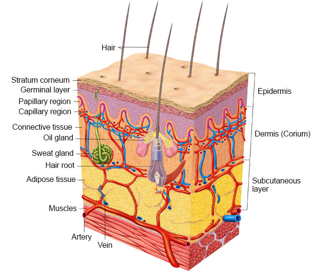
The epidermis is continuously renewed as basal cells in the lowest layer of the epidermis differentiate into keratinocytes. On their way towards the surface of the epidermis they produce keratin which gives the upper layer its typical structure with cornification. The uppermost layer (stratum corneum) consists of dead keratinocytes, which together with lipids and ceramides form a layer that is almost impermeable to water. The dead cells are constantly shed off and replaced by new cells, a process which takes about 4 weeks in humans [Iismaa et al. 2018]. In addition, melanocytes within the epidermis secrete melanin, a pigment responsible for the color of the skin, and Langerhans cells participate in the immune defense [Pérez-Sánchez et al. 2018].
The dermis is found below the epidermis, separated by the basal membrane which is mainly composed of a net of collagen fibrils (collagen I and III). Together with other proteins such as elastin and proteoglycans, the basal membrane forms the extracellular matrix (ECM) of the skin [Pérez-Sánchez et al. 2018, Sibilla et al. 2015]. The dermis is responsible for the mechanical properties of the skin (e.g., structure, elasticity and strength) and determines the outer appearance of the body. Fibroblasts producing ECM proteins and immune cells are also found in the dermis. In contrast to the epidermis, the dermis is well supplied with blood and thus provides nutrients for both itself and the epidermis. In addition, lymph vessels, hair follicles, sweat glands, and sensory cells for heat, cold, and mechanical stimuli are found within the dermis [Pérez-Sánchez et al. 2018].
The subcutis, or subcutaneous fat tissue layer, consists of fat cells that form an insulating layer against heat and cold, provide protective padding, and serve as an energy reservoir [Pérez-Sánchez et al. 2018].
The Aging process of the Skin
As we age, the composition of cells and ECM in the skin change. This results in wrinkles, uneven pigmentation, rough skin areas, loss of tension and an impaired wound healing. Aging is influenced by many factors such as genetics and hormones (intrinsic aging), and environmental factors such as UV radiation, smoking, alcohol consumption, skin disease, stress, and nutrition (extrinsic factors) [Pérez-Sánchez et al. 2018, Spiro, Lockyer 2018, Sibilla et al. 2015]. These different factors could have a cumulative effect [Spiro, Lockyer 2018].
With increasing age, the body’s ability to synthesize collagen decreases by approximately 1.5% per year, resulting primarily in a loss of collagen type I and the fragmentation of macromolecules [Sibilla et al. 2015, Cole et al. 2018]. The accumulation of collagen fragments affects the remodeling of ECM and is considered a crucial determinant in skin aging [Fisher et al. 2008]. When food is metabolized, sugars are broken down into glucose and fructose, which enter the skin via the blood and react with protein molecules of the skin. This results in cross-linking of collagen or elastin and the formation of so-called AGEs (advanced glycation end products), which lead to increased stiffness and reduced elasticity of the ECM [Clatici et al. 2017, Cole et al. 2018].
All of these changes affect the communication between the matrix and fibroblasts, promoting aging of the cells. In addition, inflammatory mediators bound to the matrix are released and accelerate aging. UV radiation and the resulting oxidizing substances have an even more profound impact on the skin [Cole et al. 2018]. Restoring the biological function of ECM may prevent the effects of aging and age-related pathological skin reactions, and contribute to health in general [Cole et al. 2018].
Effects of Bioactive Collagen Peptides on the Skin
Various studies have shown an impact of Bioactive Collagen Peptides on fibroblasts and keratinocytes, as well as on the stimulation of collagen type I and glucosaminoglycan production [Sibilla et al. 2015].
For example, Zague et al. investigated the influence of Bioactive Collagen Peptides compared to casein on the skin of rats for four weeks. Notably, protein intake was higher in the diet of the casein group than in the collagen peptide group, and collagen is known to have a lower biological value than casein. Nevertheless, skin collagen type I and IV were found to be increased in a statistically significant way in the Bioactive Collagen Peptides group, and matrix metalloproteinase MMP-2 was decrease in a statistically significant way. MMP-2 is known to primarily degrade collagen type IV, which is an important protein of the basal membrane. Its deficiency can promote the formation of wrinkles. MMP-9 is another enzyme that degrades collagen fragments and thus contributes to the remodeling of ECM. However, MMP-9 was unaffected by the intake of Bioactive Collagen Peptides [Zague et al. 2011].
Another study investigated effects on the epidermis and hair follicles using mice and cell culture models [Le Vu 2015]. Bioactive Collagen Peptides and Pro-Hyp, a dipeptide that can be detected in the blood after the ingestion of Bioactive Collagen Peptides, were analyzed. The study revealed an effect not only on the development of keratinocytes, but also on the growth of hair. This effect may be important in wound healing as well as in the protective effect of the skin against UV radiation.
Another study analyzed the effect of Bioactive Collagen Peptides on fibroblasts in cell culture mimicking sun-exposed and sun-protected human skin [Zague et al. 2017]. The authors showed that Bioactive Collagen Peptides modulate cellular metabolism of fibroblasts without affecting cell growth. Fibroblasts derived from sun-exposed skin reacted with an increased collagen synthesis even at a low concentration of collagen peptides.
Animal experiments could help clarify the mechanisms of action, however, results cannot easily be transferred to humans, as skin structure, enzymatic activity, and metabolism largely differ between species [Spiro et al. 2018].
Clinical Studies
In a study of 69 women aged 35 to 55, the effect of a specific preparation of Bioactive Collagen Peptides given in two different doses (2.5 g and 5 g daily) was tested against a placebo. Skin elasticity, skin hydration, transepidermal water loss, and skin surface texture were measured on the inside of the forearm at baseline, after 4 and 8 weeks, and 4 weeks after finishing the administration [Proksch et al. 2014a]. Both peptide doses resulted in a statistically significant increase in skin elasticity after four and eight weeks, while no statistically significant improvement was observed for the remaining parameters. Notably, a subgroup analysis showed that the effects were much more pronounced in the 50+ group than in younger participants.
The influence on the characteristics of wrinkles, and the formation of procollagen I, elastin, and fibrillin in the dermis was investigated by the same author in 57 women aged 45 to 65 and compared to a placebo [Proksch et al. 2014b]. The volunteers received 2.5 g of Bioactive Collagen Peptides or placebo for 8 weeks. The eye wrinkle volume (so-called crow’s feet wrinkles) was measured with an objective method at baseline, after 4 and 8 weeks, and 4 weeks after finishing the administration. Already by 4 weeks, the wrinkle volume showed a statistically significant decrease compared to baseline. After 8 weeks, the average decrease was 17.7% in the verum group, while the placebo group experienced an increase of 14.5%. At this point of time, the amount of of procollagen I, elastin, and fibrillin within the tissue was also determined compared to baseline. The increase in procollagen I and elastin was statistically significant at 65% and 18%, respectively, after 8 weeks. Fibrillin showed an increase of 6%, although no statistical significance was found. To summarize the study, the authors found a direct effect of Bioactive Collagen Peptides on the skin, which was stable for 4 weeks after the end of administration [Proksch et al. 2014b].
Another study compared the effect of pig collagen peptides, fish collagen peptides (both 1 0g daily), and a placebo with respect to skin moisture [Asserin et al. 2015]. After the 8-week administration, both peptide preparations had increased skin hydration, although the results were statistically significant only for pig collagen. The authors did not find an effect on transepidermal water loss. The same study also investigated the effect of fish collagen on the fragmentation of skin collagen. Here, 10 g collagen peptides daily for two weeks reduced collagen fragmentation. However, since only two volunteers were evaluated, and the difference to placebo was not statistically significant, the relevance is not clear [Spiro et al. 2018].
In summary, Bioactive Collagen Peptides may be beneficial to improve skin texture. There is some evidence of an increased effect in older women. However, the impact of ethnic diversity has not been analyzed so far. In addition, no study compared the application of collagen peptides with other factors, such as the application of sun protection or variations in the diet. Certainly, when Bioactive Collagen Peptides are applied to prevent premature skin aging, a healthy lifestyle and sufficient protection of the skin against UV radiation have to be ensured as well.
Last update: May 2022